Monday, December 27, 2010

Carcinoid tumor and Carcinoid syndrome


       Carcinoid tumor
            A carcinoid tumor is a tumor that develops from enterochromaffin cells. Enterochromaffin cells are hormone-producing cells that normally are found in the small intestine, appendix, colon, rectum, bronchi, pancreas, ovaries, testes, bile ducts, liver, as well as other organs. Enterochromaffin cells produce many types of hormones for example, histamine, serotonin, dopamine, tachykinins, and other substances that have profound effects on the circulatory system (heart and blood vessels), the gastrointestinal tract, and the lungs. For example, serotonin can cause diarrhea, histamine wheezing, and tachykinins flushing due to dilation of blood vessels.
Since carcinoid tumors develop from enterochromaffin cells, they frequently retain the capability of producing the same hormones, often in large quantities. When these hormones circulate in the blood, they can cause symptoms of carcinoid syndrome, which is discussed later.
The important characteristic of carcinoid tumors that sets them apart from other gastrointestinal tract tumors is their potential to cause the carcinoid syndrome. Most other gastrointestinal tract tumors (such as colon cancers or small bowel lymphomas) cause symptoms primarily due to their local effects on the intestines such as abdominal pain, intestinal bleeding, and intestinal obstruction. Although carcinoid tumors may also cause these local symptoms, they may also produce and release hormones that cause the carcinoid syndrome. Often, symptoms of the carcinoid syndrome can be more devastating than the local symptoms.


 Carcinoid syndrome
The carcinoid syndrome is a combination of symptoms caused by the hormones released by the tumors into the blood stream. The symptoms of the carcinoid syndrome vary depending on which hormones are released by the tumors. The common hormones released are serotonin, bradykinin (a molecule produced by enzymes at the site of an injury and then binds to receptors to cause pain), histamine, and chromogranin A (a general marker for neuroendocrine tumors). Typical carcinoid symptoms include:
• flushing
• diarrhea
• abdominal pain
and wheezing due to bronchospasm (airway narrowing)
valvular heart disease
• surgery can provoke a complication known as carcinoid crisis.
Flushing
Flushing is the most common symptom of carcinoid syndrome. An estimated 90% of the patients have flushing some time during their illness. Flushing is characterized by redness or purple discoloration of face and neck (or upper body) accompanied by a warm sensation. Episodes of flushing typically occur suddenly, either spontaneously or brought on by emotional stress, physical stress, or drinking alcohol. Episodes of flushing can last minutes to hours. Flushing can be accompanied by palpitations, low blood pressure, or fainting if blood pressure becomes too low to supply blood to the brain. Rarely, flushing can be accompanied by high blood pressure. Hormones responsible for flushing have not been clearly identified; possible candidates include serotonin, bradykinin, and substance P.
Diarrhea
Diarrhea is the second most common symptom of carcinoid syndrome. An estimated 75% of patients with carcinoid syndrome have diarrhea. Diarrhea often occurs with flushing but also can occur alone. Diarrhea in the carcinoid syndrome is most likely caused by the hormone serotonin. Medications that block the action of serotonin such as ondansetron (Zofran) often alleviates the diarrhea. Sometimes diarrhea in the carcinoid syndrome can be due to a local effect of the tumor obstructing the small intestine.
Heart disease
Heart disease occurs in an estimated 50% of patients with the carcinoid syndrome. Carcinoid syndrome typically causes scarring and stiffness of the tricuspid and pulmonic valves of the right side of the heart. Stiffness of these two valves decreases the ability of the heart to pump blood from the right ventricle to the lungs and to the rest of the body and leads to heart failure. Typical symptoms of heart failure include an enlarged liver (due to the backup of blood returning to the failing heart which is unable to pump all of the blood returning to it), swelling of the feet and ankles (edema), and swelling of the abdomen due to fluid accumulation (ascites). The damage to the tricuspid and pulmonic valves of the heart in the carcinoid syndrome is most likely caused by prolonged exposure to high levels of serotonin in the blood.
Carcinoid crisis
Carcinoid crisis is a dangerous condition that can occur at the time of surgery. It is characterized by a sudden and profound drop in blood pressure causing shock, sometimes accompanied by an abnormally fast heart rate, high blood glucose, and severe bronchospasm. Carcinoid crisis can be fatal. The best way to prevent carcinoid crisis is to treat patients undergoing surgery with somatostatin (see below) before surgery begins.
Wheezing
Wheezing occurs in approximately 10% of the patients with carcinoid syndrome. It is a result of bronchospasm (constriction of the bronchial airways) caused by hormones released by the carcinoid tumors.
Abdominal pain
Abdominal pain is common in patients with carcinoid syndrome. The pain may be due to tumor metastases in the liver, tumor invading neighboring tissues and organs.

Adenoma


       An adenoma is a benign tumor that has a glandular origin. Adenomas typically originate from cells used for secretion. Called epithelial cells, these cells are located throughout the body. However, only a portion of such cells is used for secretion. An adenoma forms when normal epithelial cells grow excessively.
Epithelial cells that are used for secretion make up specific parts of the body referred to as glands. Glands have the job of forming a number of substances in the body including, but not limited to, sweat, saliva, breast milk, mucous, and hormones. An adenoma can form from just about any glandular cell in the body.
An adenoma may form in a similar way to a malignant or cancerous tumor. A major difference between malignant tumors and adenomas is that adenomas do not metastasize or spread to other organs or tissues. Sometimes, however, adenomas develop into malignancies. When this happens, the newly malignant adenoma is called an adenocarcinoma.
Though it is possible for some adenomas to develop into cancerous tumors, having an adenoma is not usually predictive of the development of a malignancy. Most adenomas remain benign and do not form carcinomas. However, colon and rectal cancers may begin as adenomas. Additionally, bronchial adenomas can develop into lung cancer.
Frequently, adenomas have a noticeable affect on the organs or gland tissue in which they develop. Often, adenomas secrete hormones. When this occurs, the effects can be quite uncomfortable for the affected individual. In certain situations, the effects can even be deadly. However, some adenomas develop without any demonstrable effects.
There are certain types of adenomas that are more common in women, such as adenomas of the liver. Others, such as colon adenomas, are most common in adults of advancing age. The cause of adenomas is not yet known.
There are some things that seem to make developing adenomas more likely. For example, women who use oral contraceptives may be at increased risk of developing liver adenomas. Furthermore, certain types of adenomas may be inheritable. This is true of colon adenomas.
Symptoms related to adenomas vary widely. For example, a breast adenoma, called a fibroadenoma, typically causes no symptoms and may be so small that the affected individual is unable to detect it. Other breast adenomas, however, may be large enough to be noticeable by touch. By contrast, a lung adenoma can cause fever, chills, shortness of breath, and a bloody cough.
Pituitary Adenoma
• Patients tend to recover more quickly, and experience fewer complications than with conventional methods. During endoscopic pituitary adenoma surgery, access to the tumor is created via a small cut made at the rear of the nasal cavity.
• People may want to get second or third opinions prior to determining what to do if they’re diagnosed with a pituitary adenoma. It’s also challenging to discuss treatment and survival rate. A high percentage of people with an adenoma will receive excellent care and live to a ripe age.
Adenoma Tumors
• Unfortunately, the risk of the tumor becoming malignant is not reduced even if the use of steroids or contraceptives is stopped. Hepatic adenoma tumors that cause symptoms and tumors larger than 2 inches (5 cm) in size are almost always removed via surgery.
• Occasionally, adenomas begin to bleed spontaneously, and this might prove life-threatening without surgery. In the main, though, an adenoma is benign and won’t cause cancerous tumors to develop elsewhere, so outlook with treatment is good.
Kidney Adenoma
• The condition cannot be cured, and treatment measures are generally aimed at reducing symptoms and the chances of dangerous medical complications. Adenoma sebaceum tends to emerge around the age of two and worsen until a child reaches puberty.
• What type of kidney mass a patient has is generally determined by doing a biopsy and each tumor is classified by the location and nature of the mass. Renal adenoma is the most common form of benign kidney mass. Renal adenomas generally present themselves as small growths on either kidney and are mostly asymptomatic.
Adenoma Symptoms
• Another factor that might influence the development of these tumors is an imbalance in insulin and glucagons, the hormones that balance blood sugar levels and blood sugar uptake by cells. The most common hepatic adenoma symptom is pain in the upper right portion of the abdomen. As much as 50 percent of people with this tumor will experience pain in this region.
• People may want to get second or third opinions prior to determining what to do if they’re diagnosed with a pituitary adenoma. It’s also challenging to discuss treatment and survival rate. A high percentage of people with an adenoma will receive excellent care and live to a ripe age.

Crohn's disease

Crohn's disease
Crohn's disease (also spelled Crohn disease) is a chronic inflammatory disease of the intestines. It primarily causes ulcerations (breaks in the lining) of the small and large intestines, but can affect the digestive system anywhere from the mouth to the anus. It is named after the physician who described the disease in 1932. It also is called granulomatous enteritis or colitis, regional enteritis, ileitis, or terminal ileitis.
Crohn's disease is related closely to another chronic inflammatory condition that involves only the colon called ulcerative colitis. Together, Crohn's disease and ulcerative colitis are frequently referred to as inflammatory bowel disease (IBD). Ulcerative colitis and Crohn's disease have no medical cure. Once the diseases begin, they tend to fluctuate between periods of inactivity (remission) and activity (relapse).
Inflammatory bowel disease affects approximately 500,000 to two million people in the United States. Men and women are affected equally. Americans of Jewish European descent are 4 to 5 times more likely to develop IBD than the general population. IBD has historically been considered predominately disease of Caucasians, but there has been an increase in reported cases in African Americans suffering from IBD. The prevalence appears to be lower among Hispanic and Asian populations. IBD most commonly begins during adolescence and early adulthood (usually between the ages of 15 and 35). There is a small second peak of newly-diagnosed cases after age 50. The number of new cases (incidence) and number of cases (prevalence) of Crohn's disease in the United States are rising, although the reason for this is not completely understood.
Crohn's disease tends to be more common in relatives of patients with Crohn's disease. If a person has a relative with the disease, his/her risk of developing the disease is estimated to be at least 10 times that of the general population and 30 times greater if the relative with Crohn's disease is a sibling. It also is more common among relatives of patients with ulcerative colitis.

 Symptoms 
Common symptoms of Crohn's disease include abdominal pain, diarrhea, and weight loss. Less common symptoms include poor appetite, fever, night sweats, rectal pain, and occasionally rectal bleeding. The symptoms of Crohn's disease are dependent on the location, the extent, and the severity of the inflammation. The different subtypes of Crohn's disease and their symptoms are:
1. Crohn's colitis is inflammation that is confined to the colon. Abdominal pain and bloody diarrhea are the common symptoms. Anal fistulae and peri-rectal abscesses also can occur.
2. Crohn's enteritis refers to inflammation confined to the small intestine (the first part, called the jejunum or the second part, called the ileum). Involvement of the ileum alone is referred to as Crohn's ileitis. Abdominal pain and diarrhea are the common symptoms. Obstruction of the small intestine also can occur.
3. Crohn's terminal ileitis is inflammation that affects only the very end of the small intestine (terminal ileum), the part of the small intestine closest to the colon. Abdominal pain and diarrhea are the common symptoms. Small intestinal obstruction also can occur.
4. Crohn's entero-colitis and ileo-colitis are terms to describe inflammation that involve both the small intestine and the colon. Bloody diarrhea and abdominal pain are the common symptoms. Small intestinal obstruction also can occur.
Crohn's terminal ileitis and ileo-colitis are the most common types of Crohn's disease. (Ulcerative colitis frequently involves only the rectum or rectum and sigmoid colon at the distal end of the colon. These are called ulcerative proctitis and procto-sigmoiditis, respectively.)
Up to one-third of patients with Crohn's disease may have one or more of the following conditions involving the anal area:
1. Swelling of the tissue of the anal sphincter, the muscle at the end of the colon that controls defecation.
2. Development of ulcers and fissures (long ulcers) within the anal sphincter. These ulcers and fissures can cause bleeding and pain with defecation.
3. Development of anal fistulae (abnormal tunnels) between the anus or rectum and the skin surrounding the anus). Mucous and pus may drain from the openings of the fistulae on the skin.

4. Development of peri-rectal abscesses (collections of pus in the anal and rectal area). Peri-rectal abscesses can cause fever, pain and tenderness around the anus.
 Treatment
The symptoms and severity of Crohn's disease vary among patients. Patients with mild or no symptoms may not need treatment. Patients whose disease is in remission (where symptoms are absent) also may not need treatment.
Crohn's disease medications
There is no medication that can cure Crohn's disease. Patients with Crohn's disease typically will experience periods of relapse (worsening of inflammation) followed by periods of remission (lessening of inflammation) lasting months to years. During relapses, symptoms of abdominal pain, diarrhea, and rectal bleeding worsen. During remissions, these symptoms improve. Remissions usually occur because of treatment with medications or surgery, but occasionally they occur spontaneously without any treatment.
Since there is no cure for Crohn's disease, the goals of treatment are to 1) induce remissions, 2) maintain remissions, 3) minimize side effects of treatment, and 4) improve the quality of life. Treatment of Crohn's disease and ulcerative colitis with medications is similar though not always identical.
Medications for treating Crohn's disease include
1. anti-inflammatory agents such as 5-ASA compounds and corticosteroids,
2. topical antibiotics, and
3. immuno-modulators.
Selection of treatment regimens depends on disease severity, disease location, and disease-associated complications. Various guidelines recommend that approaches be sequential - initially to induce clinical remission, and then to maintain remissions. Initial evidence of improvement should be seen within 2 to 4 weeks and maximal improvement should be seen in 12 to 16 weeks3. The classic approach to therapy in Crohn's disease has been a "step-up" approach starting with the least toxic agents for mild disease, and increasingly more aggressive treatment for more severe disease, or patients who have not responded to less toxic agents. More recently the field has been moving toward a "top-down" approach (early aggressive management) which might decrease exposure to anti-inflammatory agents and increase exposure to agents that enhance mucosal healing that might prevent future complications.

Appendix


Appendix
The appendix is a closed-ended, narrow tube up to several inches in length that attaches to the cecum (the first part of the colon) like a worm. (The anatomical name for the appendix, vermiform appendix, means worm-like appendage.) The inner lining of the appendix produces a small amount of mucus that flows through the open center of the appendix and into the cecum. The wall of the appendix contains lymphatic tissue that is part of the immune system for making antibodies. Like the rest of the colon, the wall of the appendix also contains a layer of muscle, but the layer of muscle is poorly developed.


Appendicitis and what causes appendicitis
Appendicitis means inflammation of the appendix. It is thought that appendicitis begins when the opening from the appendix into the cecum becomes blocked. The blockage may be due to a build-up of thick mucus within the appendix or to stool that enters the appendix from the cecum. The mucus or stool hardens, becomes rock-like, and blocks the opening. This rock is called a fecalith (literally, a rock of stool). At other times, the lymphatic tissue in the appendix might swell and block the appendix. After the blockage occurs, bacteria which normally are found within the appendix begin to invade (infect) the wall of the appendix. The body responds to the invasion by mounting an attack on the bacteria, an attack called inflammation. An alternative theory for the cause of appendicitis is an initial rupture of the appendix followed by spread of bacteria outside of the appendix. The cause of such a rupture is unclear, but it may relate to changes that occur in the lymphatic tissue, for example, inflammation, that lines the wall of the appendix.)
If the inflammation and infection spread through the wall of the appendix, the appendix can rupture. After rupture, infection can spread throughout the abdomen; however, it usually is confined to a small area surrounding the appendix (forming a peri-appendiceal abscess).
Sometimes, the body is successful in containing ("healing") the appendicitis without surgical treatment if the infection and accompanying inflammation do not spread throughout the abdomen. The inflammation, pain and symptoms may disappear. This is particularly true in elderly patients and when antibiotics are used. The patients then may come to the doctor long after the episode of appendicitis with a lump or a mass in the right lower abdomen that is due to the scarring that occurs during healing. This lump might raise the suspicion of cancer.

 Symptoms 
The main symptom of appendicitis is abdominal pain. The pain is at first diffuse and poorly localized, that is, not confined to one spot. (Poorly localized pain is typical whenever a problem is confined to the small intestine or colon, including the appendix.) The pain is so difficult to pinpoint that when asked to point to the area of the pain, most people indicate the location of the pain with a circular motion of their hand around the central part of their abdomen. A second, common, early symptom of appendicitis is loss of appetite which may progress to nausea and even vomiting. Nausea and vomiting also may occur later due to intestinal obstruction.
As appendiceal inflammation increases, it extends through the appendix to its outer covering and then to the lining of the abdomen, a thin membrane called the peritoneum. Once the peritoneum becomes inflamed, the pain changes and then can be localized clearly to one small area. Generally, this area is between the front of the right hip bone and the belly button. The exact point is named after Dr. Charles McBurney--McBurney's point. If the appendix ruptures and infection spreads throughout the abdomen, the pain becomes diffuse again as the entire lining of the abdomen becomes inflamed.


 Treatment
Once a diagnosis of appendicitis is made, an appendectomy usually is performed. Antibiotics almost always are begun prior to surgery and as soon as appendicitis is suspected.
There is a small group of patients in whom the inflammation and infection of appendicitis remain mild and localized to a small area. The body is able not only to contain the inflammation and infection but to resolve it as well. These patients usually are not very ill and improve during several days of observation. This type of appendicitis is referred to as "confined appendicitis" and may be treated with antibiotics alone. The appendix may or may not be removed at a later time.
On occasion, a person may not see their doctor until appendicitis with rupture has been present for many days or even weeks. In this situation, an abscess usually has formed, and the appendiceal perforation may have closed over. If the abscess is small, it initially can be treated with antibiotics; however, the abscess usually requires drainage. A drain (a small plastic or rubber tube) usually is inserted through the skin and into the abscess with the aid of an ultrasound or CT scan that can determine the exact location of the abscess. The drain allows pus to flow from the abscess out of the body. The appendix may be removed several weeks or months after the abscess has resolved. This is called an interval appendectomy and is done to prevent a second attack of appendicitis.

Gastritis


Gastritis
  Gastritis is a condition in which the stomach lining—known as the mucosa—is inflamed. The stomach lining contains special cells that produce acid and enzymes, which help break down food for digestion, and mucus, which protects the stomach lining from acid. When the stomach lining is inflamed, it produces less acid, enzymes, and mucus.
Gastritis may be acute or chronic. Sudden, severe inflammation of the stomach lining is called acute gastritis. Inflammation that lasts for a long time is called chronic gastritis. If chronic gastritis is not treated, it may last for years or even a lifetime.
Erosive gastritis is a type of gastritis that often does not cause significant inflammation but can wear away the stomach lining. Erosive gastritis can cause bleeding, erosions, or ulcers. Erosive gastritis may be acute or chronic.
The relationship between gastritis and symptoms is not clear. The term gastritis refers specifically to abnormal inflammation in the stomach lining. People who have gastritis may experience pain or discomfort in the upper abdomen, but many people with gastritis do not have any symptoms.
The term gastritis is sometimes mistakenly used to describe any symptoms of pain or discomfort in the upper abdomen. Many diseases and disorders can cause these symptoms. Most people who have upper abdominal symptoms do not have gastritis.


 Symptoms 
Many people with gastritis do not have any symptoms, but some people experience symptoms such as
• upper abdominal discomfort or pain
• nausea
• vomiting
These symptoms are also called dyspepsia.
Erosive gastritis may cause ulcers or erosions in the stomach lining that can bleed. Signs of bleeding in the stomach include
• blood in vomit
• black, tarry stools
• red blood in the stool
.
 Treatment 
Medications that reduce the amount of acid in the stomach can relieve symptoms that may accompany gastritis and promote healing of the stomach lining. These medications include
• antacids, such as aspirin, sodium bicarbonate, and citric acid (Alka-Seltzer); alumina and magnesia (Maalox); and calcium carbonate and magnesia (Rolaids). Antacids relieve mild heartburn or dyspepsia by neutralizing acid in the stomach. These drugs may produce side effects such as diarrhea or constipation.
• histamine 2 (H2) blockers, such as famotidine (Pepcid AC) and ranitidine (Zantac 75). H2 blockers decrease acid production. They are available both over the counter and by prescription.
• proton pump inhibitors (PPIs), such as omeprazole (Prilosec, Zegerid), lansoprazole (Prevacid), pantoprazole (Protonix), rabeprazole (Aciphex), esomeprazole (Nexium), and dexlansoprazole (Kapidex). All of these drugs are available by prescription, and some are also available over the counter. PPIs decrease acid production more effectively than H2 blockers.
Depending on the cause of the gastritis, additional measures or treatments may be needed. For example, if gastritis is caused by prolonged use of NSAIDs, a doctor may advise a person to stop taking NSAIDs, reduce the dose of NSAIDs, or switch to another class of medications for pain. PPIs may be used to prevent stress gastritis in critically ill patients.
Treating H. pylori infections is important, even if a person is not experiencing symptoms from the infection. Untreated H. pylori gastritis may lead to cancer or the development of ulcers in the stomach or small intestine. The most common treatment is a triple therapy that combines a PPI and two antibiotics—usually amoxicillin and clarithromycin—to kill the bacteria. Treatment may also include bismuth subsalicylate (Pepto-Bismol) to help kill bacteria.
After treatment, the doctor may use a breath or stool test to make sure the H. pylori infection is gone. Curing the infection can be expected to cure the gastritis and decrease the risk of other gastrointestinal diseases associated with gastritis, such as peptic ulcer disease, gastric cancer, and MALT lymphoma.

Hernia


Hernia
A hernia is an opening or weakness in the muscular structure of the wall of the abdomen. This defect causes a bulging of the abdominal wall. This bulging is usually more noticeable when the abdominal muscles are tightened, thereby increasing the pressure in the abdomen. Any activities that increase intra-abdominal pressure can worsen a hernia; examples of such activities are lifting, coughing, or even straining to have a bowel movement. Imagine a barrel with a hole in its side and a balloon that is blown up inside the barrel. Part of the inflated balloon would bulge out through the hole. The balloon going through the hole is like the tissues of the abdomen bulging through a hernia.
Serious complications from a hernia can result from the trapping of tissues in the hernia -- a process called incarceration. Trapped or incarcerated tissues may have their blood supply cut off, leading to damage or death of the tissue. The treatment of an incarceration usually involves surgery.
Where are hernias located?
The most common location for hernias is the groin (or inguinal) area. There are several reasons for this tendency. First, there is a natural anatomical weakness in the groin region which results from incomplete muscle coverage. Second, the upright position of human posture results in a greater force pushing toward the bottom of the abdomen, thereby increasing the stress on these weaker tissues. The combination of these factors over time breaks down the support tissues, enlarging any preexisting hole, or leads to a tear, resulting in a new hole.
Several different types of hernia may occur, and frequently coexist, in the groin area. These include indirect, direct, and femoral hernias, which are defined by the location of the opening of the hernia from the abdomen to the groin. Another type of hernia, called a ventral hernia, occurs in the midline of the abdomen, usually above the navel (umbilicus). This type of hernia is usually painless. Hernias can also occur within the navel (umbilical hernia).

Symptoms and signs
Symptoms of a hernia include pain or discomfort and a localized swelling somewhere on the surface of the abdomen or in the groin area.


 Anesthesia is used for hernia surgery
Most hernia repairs can be done with a variety of anesthetic methods. With modern general anesthetic techniques and monitoring, general anesthesia can be very safe. However the surgery can also be performed under local anesthesia or regional anesthetics, often using sedation medications at the same time to help relax the patient. The specific type of anesthetic for an individual patient is selected after careful evaluation of the patient's general health and individual concerns.


What can be done to prevent a hernia?
Most of the factors that lead to the development of hernias are beyond the control of the individual. Some of those factors are inherited and develop as the individual grows. The arrangement of the local tissues and their thickness and strength may greatly affect the relative risk of developing a hernia over a lifetime. However, that risk can be increased by failure to use good body mechanics when lifting, poor abdominal support posture, and weight-control problems.


Are hernias inherited?
Since genetics dictate inherited features and structure, there is a significant risk of inheriting the anatomical features that may predispose to a hernia. There may also be inherited factors that result in tissue weakness, which ultimately allows the deterioration of the supporting structures and leads to the formation of a hernia. However, this does not necessarily imply that the offspring of an individual with a hernia will ultimately develop the problem.


Do hernias usually develop on both sides of the body?
Groin hernias are somewhat more likely to develop on both sides. This is probably because the structural elements develop symmetrically, and the stresses on the body that occur over time are similar on both sides. When a patient becomes aware of a groin swelling on one side, examination by a doctor will often identify a small hernia on the opposite side.

Achalasia

 Achalasia
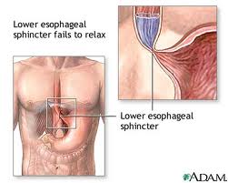
Achalasia is a rare disease of the muscle of the esophagus (swallowing tube). The term achalasia means "failure to relax" and refers to the inability of the lower esophageal sphincter (a ring of muscle situated between the lower esophagus and the stomach) to open and let food pass into the stomach. As a result, patients with achalasia have difficulty in swallowing food.


 Causes 
The cause of achalasia is unknown. Theories on causation invoke infection, heredity or an abnormality of the immune system that causes the body itself to damage the esophagus (autoimmune disease).
The esophagus contains both muscles and nerves. The nerves coordinate the relaxation and opening of the sphincters as well as the peristaltic waves in the body of the esophagus. Achalasia has effects on both the muscles and nerves of the esophagus; however, the effects on the nerves are believed to be the most important. Early in achalasia, inflammation can be seen (when examined under the microscope) in the muscle of the lower esophagus, especially around the nerves. As the disease progresses, the nerves begin to degenerate and ultimately disappear, particularly the nerves that cause the lower esophageal sphincter to relax. Still later in the progression of the disease, muscle cells begin to degenerate, possibly because of the damage to the nerves. The result of these changes is a lower sphincter that cannot relax and muscle in the lower esophageal body that cannot support peristaltic waves. With time, the body of the esophagus stretches and becomes enlarged (dilated).

Symptoms 
The most common symptom of achalasia is difficulty in swallowing (dysphagia). Patients typically describe food sticking in the chest after it is swallowed. Dysphagia occurs with both solid and liquid food. Moreover, the dysphagia is consistent, meaning that it occurs during virtually every meal.
Sometimes, patients will describe a heavy sensation in their chest after eating that may force them to stop eating. Occasionally, pain may be severe and mimic heart pain (angina). The cause of this discomfort is felt to be the accumulation of ingested food within the esophagus.
Regurgitation of food that is trapped in the esophagus can occur, especially when the esophagus is dilated. If the regurgitation happens at night while the patient is sleeping, food can enter the throat and cause coughing and choking. If the food enters the trachea (windpipe) and lung, it can lead to infection (aspiration pneumonia).
Because of the problem with swallowing food, a large proportion of patients with achalasia lose weight. Episodes of chest pain may also occur especially with vigorous achalasia. Sometimes symptoms suggest gastroesophageal reflux disease (GERD); however, it's not clear if the symptoms are, in fact, due to reflux. Moreover, acid suppression rarely improves the symptoms of achalasia.

 Treatment
Treatments for achalasia include oral medications, stretching of the lower esophageal sphincter (dilation), surgery to cut the sphincter (esophagomyotomy), and the injection of botulinum toxin (Botox) into the sphincter. All four treatments reduce the pressure within the lower esophageal sphincter to allow easier passage of food from the esophagus into the stomach.
Oral medications
Oral medications that help to relax the lower esophageal sphincter include groups of drugs called nitrates, for example, isosorbide dinitrate (Isordil) and calcium channel blockers, for example, nifedipine (Procardia) and verapamil (Calan). Although some patients with achalasia, particularly early in the disease, have improvement of symptoms with medications, most do not. By themselves, oral medications are likely to provide only short-term and not long-term relief of the symptoms of achalasia, and many patients experience side-effects from the medications.
Dilation
The lower esophageal sphincter also may be treated directly by forceful dilation. Dilation of the lower esophageal sphincter is done by having the patient swallow a tube with a balloon at the end. The balloon is placed across the lower sphincter with the help of X-rays, and the balloon is blown up suddenly. The goal is to stretch--actually to tear--the sphincter. The success of forceful dilation has been reported to be between 60% and 95%. Patients in whom dilation is not successful can undergo further dilations, but the rate of success decreases with each additional dilation. If dilation is not successful, the sphincter may still be treated surgically. The main complication of forceful dilation is rupture of the esophagus, which occurs 5% of the time. Half of the ruptures heal without surgery, though patients with rupture who do not require surgery should be followed closely and treated with antibiotics. The other half of ruptures require surgery. (Although surgery carries additional risk for the patient, surgery can repair the rupture as well as permanently treat the achalasia with esophagomyotomy.) Death following forceful dilation is rare. Dilation is a quick and inexpensive procedure compared with surgery, and requires only a short hospital stay.
Esophagomyotomy
The sphincter also can be cut surgically, a procedure called esophagomyotomy. The surgery can be done using an abdominal incision or laparoscopically through small punctures in the abdomen. In general, the laparoscopic approach is used with uncomplicated achalasia. Alternatively, the surgery can be done with a large incision or laparoscopically through the chest. Esophagomyotomy is more successful than forceful dilation, probably because the pressure in the lower sphincter is reduced to a greater extent and more reliably; 80%-90% of patients have good results. With prolonged follow-up, however, some patients develop recurrent dysphagia. Thus, esophagomyotomy does not guarantee a permanent cure. The most important side effect from the more reliable and greater reduction in pressure with esophagomyotomy, is reflux of acid (gastroesophageal reflux disease or GERD). In order to prevent this, the esophagomyotomy can be modified so that it doesn't completely cut the sphincter or the esophagomyotomy may be combined with anti-reflux surgery (fundoplication). Whichever surgical procedure is done, some physicians recommend life-long treatment with oral medications for acid reflux. Others recommend 24 hour esophageal acid testing with lifelong medication only if acid reflux is found.

Sunday, December 26, 2010

Jaundice


         Jaundice is not a disease but rather a sign that can occur in many different diseases. Jaundice is the yellowish staining of the skin and sclerae (the whites of the eyes) that is caused by high levels in blood of the chemical bilirubin. The color of the skin and sclerae vary depending on the level of bilirubin. When the bilirubin level is mildly elevated, they are yellowish. When the bilirubin level is high, they tend to be brown.

Causes 
Bilirubin comes from red blood cells. When red blood cells get old, they are destroyed. Hemoglobin, the iron-containing chemical in red blood cells that carries oxygen, is released from the destroyed red blood cells after the iron it contains is removed. The chemical that remains in the blood after the iron is removed becomes bilirubin.

The liver has many functions. One of the liver's functions is to produce and secrete bile into the intestines to help digest dietary fat. Another is to remove toxic chemicals or waste products from the blood, and bilirubin is a waste product. The liver removes bilirubin from the blood. After the bilirubin has entered the liver cells, the cells conjugate (attaching other chemicals, primarily glucuronic acid) to the bilirubin, and then secrete the bilirubin/glucuronic acid complex into bile. The complex that is secreted in bile is called conjugated bilirubin. The conjugated bilirubin is eliminated in the feces. (Bilirubin is what gives feces its brown color.) Conjugated bilirubin is distinguished from the bilirubin that is released from the red blood cells and not yet removed from the blood which is termed unconjugated bilirubin.

Jaundice occurs when there is 1) too much bilirubin being produced for the liver to remove from the blood. (For example, patients with hemolytic anemia have an abnormally rapid rate of destruction of their red blood cells that releases large amounts of bilirubin into the blood), 2) a defect in the liver that prevents bilirubin from being removed from the blood, converted to bilirubin/glucuronic acid (conjugated) or secreted in bile, or 3) blockage of the bile ducts that decreases the flow of bile and bilirubin from the liver into the intestines. (For example, the bile ducts can be blocked by cancers, gallstones, or inflammation of the bile ducts). The decreased conjugation, secretion, or flow of bile that can result in jaundice is referred to as cholestasis: however, cholestasis does not always result in jaundice.

Increased production of bilirubin
There are several uncommon conditions that give rise to over-production of bilirubin. The bilirubin in the blood in these conditions usually is only mildly elevated, and the resultant jaundice usually is mild and difficult to detect. These conditions include: 1) rapid destruction of red blood cells (referred to as hemolysis), 2) a defect in the formation of red blood cells that leads to the over-production of hemoglobin in the bone marrow (called ineffective erythropoiesis), or 3) absorption of large amounts of hemoglobin when there has been much bleeding into tissues (e.g., from hematomas, collections of blood in the tissues).
Acute inflammation of the liver
Any condition in which the liver becomes inflamed can reduce the ability of the liver to conjugate (attach glucuronic acid to) and secrete bilirubin. Common examples include acute viral hepatitis, alcoholic hepatitis, and Tylenol-induced liver toxicity.
Chronic liver diseases
Chronic inflammation of the liver can lead to scarring and cirrhosis, and can ultimately result in jaundice. Common examples include chronic hepatitis B and C, alcoholic liver disease with cirrhosis, and autoimmune hepatitis.
Infiltrative diseases of the liver
Infiltrative diseases of the liver refer to diseases in which the liver is filled with cells or substances that don't belong there. The most common example would be metastatic cancer to the liver, usually from cancers within the abdomen. Uncommon causes include a few diseases in which substances accumulate within the liver cells, for example, iron (hemochromatosis), alpha-one antitrypsin (alpha-one antitrypsin deficiency), and copper (Wilson's disease).
Inflammation of the bile ducts
Diseases causing inflammation of the bile ducts, for example, primary biliary cirrhosis or sclerosing cholangitis and some drugs, can stop the flow of bile and elimination of bilirubin and lead to jaundice.
Blockage of the bile ducts
The most common causes of blockage of the bile ducts are gallstones and pancreatic cancer. Less common causes include cancers of the liver and bile ducts.
Drugs
Many drugs can cause jaundice and/or cholestasis. Some drugs can cause liver inflammation (hepatitis) similar to viral hepatitis. Other drugs can cause inflammation of the bile ducts, resulting in cholestasis and/or jaundice. Drugs also may interfere directly with the chemical processes within the cells of the liver and bile ducts that are responsible for the formation and secretion of bile to the intestine. As a result, the constituents of bile, including bilirubin, are retained in the body. The best example of a drug that causes this latter type of cholestasis and jaundice is estrogen. The primary treatment for jaundice caused by drugs is discontinuation of the drug. Almost always the bilirubin levels will return to normal within a few weeks, though in a few cases it may take several months.
Genetic disorders
There are several rare genetic disorders present from birth that give rise to jaundice. Crigler-Najjar syndrome is caused by a defect in the conjugation of bilirubin in the liver due to a reduction or absence of the enzyme responsible for conjugating the glucuronic acid to bilirubin. Dubin-Johnson and Rotor's syndromes are caused by abnormal secretion of bilirubin into bile.
The only common genetic disorder that may cause jaundice is Gilbert's syndrome which affects approximately 7% of the population. Gilbert's syndrome is caused by a mild reduction in the activity of the enzyme responsible for conjugating the glucuronic acid to bilirubin. The increase in bilirubin in the blood usually is mild and infrequently reaches levels that cause jaundice. Gilbert's syndrome is a benign condition that does not cause health problems.


Treatment
With the exception of the treatments for specific causes of jaundice mentioned previously, the treatment of jaundice usually requires a diagnosis of the specific cause of the jaundice and treatment directed at the specific cause, e.g., removal of a gallstone blocking the bile duct.

Fever


      Fever refers to an elevation in body temperature. Technically, any body temperature above the normal oral measurement of 98.6 F (37 C) or the normal rectal temperature of 99 F (37.2 C) is considered to be elevated. However, these are averages, and one's normal body temperature may actually be 1 F (0.6 C) or more above or below the average of 98.6 F. Body temperature can also vary up to 1 F (0.6 C) throughout the day.
Fever is not considered medically significant until body temperature is above 100.4 F (38 C). Anything above normal but below 100.4 F (38 C) is considered a low-grade fever. Fever serves as one of the body's natural defenses against bacteria and viruses which cannot live at a higher temperature. For that reason, low fevers should normally go untreated, unless accompanied by troubling symptoms.
Also, the body's defense mechanisms seem to work more efficiently at a higher temperature. Fever is just one part of an illness, many times no more important than the presence of other symptoms such as cough, sore throat, fatigue, joint pains or aches, chills, nausea, etc.
Fevers of 104 F (40 C) or higher demand immediate home treatment and subsequent medical attention, as they can result in delirium and convulsions, particularly in infants and children.
Fever should not be confused with hyperthermia, which is a defect in your body's response to heat (thermoregulation), which can also raise the body temperature. This is usually caused by external sources such as being in a hot environment.
How should I take a temperature for fever?
Digital thermometers can be used to measure rectal, oral, or axillary (under the armpit) temperatures. The American Academy of Pediatrics does not recommend use of mercury thermometers (glass), and they encourage parents to remove mercury thermometers from their households to prevent accidental exposure to this toxin.
Measuring an axillary (under the armpit) temperature for fever:
Axillary temperatures are not as accurate as rectal or oral measurements, and these generally measure 1 degree lower than a simultaneously obtained oral temperature.
Place the tip of the digital thermometer in your child's armpit.
• Leave in place about one minute or until you hear a beep to check a digital reading.
Measuring fever by eardrum temperature:
Tympanic (ear) thermometers must be placed correctly in your child's ear to be accurate. Too much earwax can cause the reading to be incorrect.
Eardrum temperature measurements are not accurate in small children and should not be used in children under 3 years (36 months) of age. This is especially true in infants below 3 months of age when obtaining an accurate temperature is very important.
Measuring fever by oral temperature:
People 4 years old and older can have their temperature taken with a digital thermometer under the tongue with their mouth closed.
• Clean the thermometer with soapy water or rubbing alcohol and rinse.

• Turn the thermometer on and place the tip of the thermometer as far back under the tongue as possible.

• The mouth should remain closed, as an open mouth can cause readings to be inaccurate.

• The thermometer should remain in place for about one minute or until you hear the beep. Check the digital reading.
Avoid hot or cold drinks within 15 minutes of oral temperature measurement to ensure correct readings.
Measuring fever by rectal temperature:
The American Academy of Pediatrics recommends rectal temperature measurements for children under 3 years of age, as this gives the most accurate reading of core temperature.
• Clean the thermometer with soapy water or rubbing alcohol and rinse with cool water.

• Use a small amount of lubricant, such as petroleum jelly, on the end.

• Place the child prone (belly-side down) on a firm surface, or place your child face up and bend his legs to his chest.

• After separating the buttocks, insert the thermometer approximately ½ to 1 inch into the rectum. Do not insert it too far.

• Hold the thermometer in place, loosely keeping your hand cupped around your child's bottom, and keep your fingers on the thermometer to avoid it accidently sliding further into the rectum. Keep it there for about one minute, until you hear the beep.

• Remove the thermometer, and check the digital reading.

• Label the rectal thermometer so it's not accidentally used in the mouth.
A rectal temperature will read approximately 1 degree higher than a simultaneously obtained oral temperature.
What is the treatment for a fever?
Generally, if the fever does not cause discomfort, the fever itself need not be treated. It is not necessary to awaken an adult or child to treat a fever unless instructed to do so by your health-care practitioner.
The following fever-reducing medications may be used at home:
• Acetaminophen (Tylenol and others) can be used to lower a fever. The recommended pediatric dose can be suggested by the child's health-care provider. Adults without liver disease or other health problems can take 1,000 mg (two "extra strength" tablets) every four to six hours or as directed by your physician.

• Ibuprofen (Motrin/Advil) can also be used to break a fever in patients over 6 months of age. Discuss the best dose with your doctor. For adults, 400-600 mg (two to three 200 mg tablets) can be used every six hours.

Aspirin should not be used for fever in children or adolescents. Aspirin use in children and adolescents during a viral illness (especially chickenpox and influenza, or flu) has been associated with Reye syndrome. Reye syndrome is a dangerous illness which causes prolonged vomiting, confusion, and even coma and liver failure.
An individual with a fever should be kept comfortable and not overdressed. Overdressing can cause the temperature to rise further. Tepid water (85 F [30 C]) baths are a home remedy that may help bring down a fever. Never immerse someone in ice water. This is a common misconception. Never sponge a child or an adult with alcohol; the alcohol fumes may be inhaled, causing many problems.

Pain

    Pain is a feeling triggered in the nervous system. Pain may be sharp or dull. It may come and go, or it may be constant. You may feel pain in one area of your body, such as your back, abdomen or chest or you may feel pain all over, such as when your muscles ache from the flu.
Pain can be helpful in diagnosing a problem. Without pain, you might seriously hurt yourself without knowing it, or you might not realize you have a medical problem that needs treatment. Once you take care of the problem, pain usually goes away. However, sometimes pain goes on for weeks, months or even years. This is called chronic pain. Sometimes chronic pain is due to an ongoing cause, such as cancer or arthritis. Sometimes the cause is unknown.
Fortunately, there are many ways to treat pain. Treatment varies depending on the cause of pain. Pain relievers, acupuncture and sometimes surgery are helpful.

There are 2 types of pain: acute and chronic
Acute pain doesn't last long and usually goes away as your body heals. Chronic pain lasts at least 6 months after your body has healed. Sometimes, people who have chronic pain don't know what is causing it. Along with discomfort, chronic pain can cause low self-esteem, depression and anger. It can also interfere with your daily activities.
 Treatment
Treatment of chronic pain usually involves medicines and therapy. Medicines used for chronic pain include pain relievers, antidepressants and anticonvulsants. Different types of medicines help people who have different types of pain. You usually use long-acting medicines for constant pain. Short-acting medicines treat pain that comes and goes.

Several types of therapy can help ease your pain. Physical therapy (such as stretching and strengthening activities) and low-impact exercise (such as walking, swimming or biking) can help reduce the pain. However, exercising too much or not at all can hurt chronic pain patients. Occupational therapy teaches you how to pace yourself and how to do ordinary tasks differently so you won't hurt yourself. Behavioral therapy can reduce your pain through methods that help you relax, such as meditation and yoga. It can also help decrease stress.

Lifestyle changes are an important part of treatment for chronic pain. Getting regular sleep at night and not taking daytime naps should help. Stopping smoking also helps because the nicotine in cigarettes can make some medicines less effective. Smokers also tend to have more pain than nonsmokers.

Most pain treatments will not take away all of your pain. Instead, treatment should reduce how much pain you have and how often it occurs. Talk to your doctor to learn how to best control your pain.

Diarrhea


Diarrhea is an increase in the frequency of bowel movements or a decrease in the form of stool (greater looseness of stool). Although changes in frequency of bowel movements and looseness of stools can vary independently of each other, changes often occur in both.
Diarrhea needs to be distinguished from four other conditions. Although these conditions may accompany diarrhea, they often have different causes and different treatments than diarrhea.

Treatment
Absorbents. Absorbents are compounds that absorb water. Absorbents that are taken orally bind water in the small intestine and colon and make diarrheal stools less watery. They also may bind toxic chemicals produced by bacteria that cause the small intestine to secrete fluid; however, the importance of toxin binding in reducing diarrhea is unclear.
The two main absorbents are attapulgite and polycarbophil, and they are both available without prescriptions.
Examples of products containing attapulgite are:
• Donnagel,
• Rheaban,
• Kaopectate Advanced Formula,
• Parepectolin, and
• Diasorb.
Examples of products containing polycarbophil are:
• Equalactin,
• Konsyl Fiber,
• Mitrolan, and
• Polycarb.
Equalactin is the antidiarrheal product containing attapulgite; however the laxative, Konsyl, also contains attapulgite. Attapulgite and polycarbophil remain in the intestine and, therefore, have no side effects outside of the gastrointestinal tract. They may occasionally cause constipation and bloating. One concern is that absorbents also can bind medications and interfere with their absorption into the body. For this reason, it often is recommended that medications and absorbents be taken several hours apart so that they are physically separated within the intestine.
Anti-motility medications. Anti-motility medications are drugs that relax the muscles of the small intestine and/or the colon. Relaxation results in slower flow of intestinal contents. Slower flow allows more time for water to be absorbed from the intestine and colon and reduces the water content of stool. Cramps, due to spasm of the intestinal muscles, also are relieved by the muscular relaxation.
The two main anti-motility medications are loperamide (Imodium), which is available without a prescription, and diphenoxylate (Lomotil), which requires a prescription. Both medications are related to opiates (for example, codeine ) but neither has the pain-relieving effects of opiates.
Loperamide (Imodium), though related to opiates, does not cause addiction.
Diphenoxylate is a man-made medication that at high doses can be addictive because of its opiate-like, euphoric (mood-elevating) effects. In order to prevent abuse of diphenoxylate and addiction, a second medication, atropine, is added to loperamide in Lomotil. If too much Lomotil is ingested, unpleasant side effects from too much atropine will occur.
Loperamide and diphenoxylate are safe and well-tolerated. There are some precautions, however, that should be observed.
• Anti-motility medications should not be used without a doctor's guidance to treat diarrhea caused by moderate or severe ulcerative colitis, C. difficile colitis, and intestinal infections by bacteria that invade the intestine (for example, Shigella). Their use can lead to more serious inflammation and prolong the infections.
• Diphenoxylate can cause drowsiness or dizziness, and caution should be used if driving or performing tasks that require alertness and coordination are required.
• Anti-motility medications should not be used in children younger than two years of age.
• Most unimportant, acute diarrhea should improve within 72 hours. If symptoms do not improve or if they worsen, a doctor should be consulted before continuing treatment with anti-motility medications.
Bismuth compounds. Many bismuth-containing preparations are available around the world. Bismuth subsalicylate (Pepto-Bismol) is available in the United States. It contains two potentially active ingredients, bismuth and salicylate (aspirin). It is not clear how effective bismuth compounds are, except in traveler's diarrhea and the treatment of H. pylori infection of the stomach where they have been shown to be effective. It also is not clear how bismuth subsalicylate might work. It is thought to have some antibiotic-like properties that affect bacteria that cause diarrhea. The salicylate is anti-inflammatory and could reduce secretion of water by reducing inflammation. Bismuth also might directly reduce the secretion of water by the intestine.
Pepto-Bismol is well-tolerated. Minor side effects include darkening of the stool and tongue. There are several precautions that should be observed when using Pepto-Bismol.
• Since it contains aspirin, patients who are allergic to aspirin should not take Pepto-Bismol.
• Pepto-Bismol should not be used with other aspirin-containing medications since too much aspirin may be ingested and lead to aspirin toxicity, the most common manifestation of which is ringing in the ears.
• The aspirin in Pepto-Bismol can accentuate the effects of anticoagulants, particularly warfarin (Coumadin), and lead to excessive bleeding. It also may cause abnormal bleeding in people who have a tendency to bleed because of genetic disorders or underlying diseases, for example, cirrhosis, that may cause abnormal bleeding.
• The aspirin in Pepto-Bismol can aggravate stomach and duodenal ulcer disease.
• Pepto-Bismol and aspirin-containing products should not be given to children and teenagers with chickenpox, influenza, and other viral infections because they may cause Reye's syndrome. Reye's syndrome is a serious illness affecting primarily the liver and brain that can lead to liver failure and coma, with a mortality rate of at least 20%.
• Pepto-Bismol should not be given to infants and children younger than two years of age.

Measles


   Measles (Rubeola) is a highly communicable disease characterized by fever, general malaise, sneezing, nasal congestion, brassy cough, conjunctivitis, and a maculopapular eruption over the entire body caused by the rubeola virus.
Description of Measles
While measles is usually a benign infection, encephalitis is a grave complication. Of those who develop encephalitis, about one in eight will die.
Bronchopneumonia is a serious complication of measles.
Otitis media, followed by mastoiditis, brain abscesses, or even meningitis, is not rare.
Causes and Risk Factors of Measles
Measles is most common in school-age children with outbreaks occurring in the winter and spring.
The occurrence of measles before the age of 6 months is relatively uncommon because of passively acquired maternal antibodies from the immune mother.
Symptoms of Measles
The onset is gradual with rhinitis, drowsiness, loss of appetite, gradual elevation of temperature for the first two days, when the fever may rise to 101 to 103 degrees (F).
Koplik's spots usually appear on the buccal (inner cheek) mucosa opposite the molars on the second or third day.
About the fourth day, fever usually reaches a higher elevation than previously, at times as high as 104 to 106 degrees (F). With this recurrence the rash appears.
The measles rash first appears on the face, being seen early as small maculopapular lesions that increase rapidly in size and coalesce in places, often causing a swollen, mottled appearance. The rash extends to the body and extremities, and in some areas may resemble the rash of scarlet fever.
A cough, present at this time, is due to bronchitis produced by the inflammatory condition of the mucous membranes, which undoubtedly corresponds to the rash seen on the skin.
Ordinarily, the rash lasts from 4 to 5 days; and as it subsides, the temperature declines. Consequently, five days after the appearance of the rash, the temperature should be normal or approximately normal in uncomplicated cases.
Treatment of Measles
Maintain bedrest and provide quiet activities for the child. If there is sensitivity to light, keep room darkly lit.
Remove eye secretions with warm saline or water. Encourage the child not to rub the eyes.
Administer antipruritic medication and tepid sponge baths as ordered.
A cool mist vaporizer can be used to relieve cough.
Apply antipruritic medication to prevent itching.
Isolate child until fifth day of rash.
Prevention of Measles
Generally two doses of live measles vaccine are recommended, one shot at 15 months of age, and the second shot before entering either kindergarten or first grade (or at some other age as required by law in your state).
Measles re-vaccination guidelines are as follows: Persons vaccinated with live measle vaccine before their first birthday should be considered unvaccinated, and should receive at least one dose of measles vaccine.
Immunization is recommended one time for all persons born after 1956 who lack evidence of immunity to measles (receipt of live vaccine on or after the first birthday, laboratory evidence of immunity, or a history of physician-documented measles).
A second dose of measles vaccine is recommended for young adults in settings where individuals congregate (colleges, residential settings, etc.).

Diabetes mellitus


Diabetes mellitus is a group of metabolic diseases characterized by high blood sugar (glucose) levels, that result from defects in insulin secretion, or action, or both. Diabetes mellitus, commonly referred to as diabetes (as it will be in this article) was first identified as a disease associated with "sweet urine," and excessive muscle loss in the ancient world. Elevated levels of blood glucose (hyperglycemia) lead to spillage of glucose into the urine, hence the term sweet urine.
Normally, blood glucose levels are tightly controlled by insulin, a hormone produced by the pancreas. Insulin lowers the blood glucose level. When the blood glucose elevates (for example, after eating food), insulin is released from the pancreas to normalize the glucose level. In patients with diabetes, the absence or insufficient production of insulin causes hyperglycemia. Diabetes is a chronic medical condition, meaning that although it can be controlled, it lasts a lifetime.
Impact of diabetes
Over time, diabetes can lead to blindness, kidney failure, and nerve damage. These types of damage are the result of damage to small vessels, referred to as microvascular disease. Diabetes is also an important factor in accelerating the hardening and narrowing of the arteries (atherosclerosis), leading to strokes, coronary heart disease, and other large blood vessel diseases. This is referred to as macrovascular disease. Diabetes affects approximately 17 million people (about 8% of the population) in the United States. In addition, an estimated additional 12 million people in the United States have diabetes and don't even know it.
From an economic perspective, the total annual cost of diabetes in 1997 was estimated to be 98 billion dollars in the United States. The per capita cost resulting from diabetes in 1997 amounted to $10,071.00; while healthcare costs for people without diabetes incurred a per capita cost of $2,699.00. During this same year, 13.9 million days of hospital stay were attributed to diabetes, while 30.3 million physician office visits were diabetes related. Remember, these numbers reflect only the population in the United States. Globally, the statistics are staggering.
Diabetes is the third leading cause of death in the United States after heart disease and cancer.
Causes
Insufficient production of insulin (either absolutely or relative to the body's needs), production of defective insulin (which is uncommon), or the inability of cells to use insulin properly and efficiently leads to hyperglycemia and diabetes. This latter condition affects mostly the cells of muscle and fat tissues, and results in a condition known as "insulin resistance." This is the primary problem in type 2 diabetes. The absolute lack of insulin, usually secondary to a destructive process affecting the insulin producing beta cells in the pancreas, is the main disorder in type 1 diabetes. In type 2 diabetes, there also is a steady decline of beta cells that adds to the process of elevated blood sugars. Essentially, if someone is resistant to insulin, the body can, to some degree, increase production of insulin and overcome the level of resistance. After time, if production decreases and insulin cannot be released as vigorously, hyperglycemia develops.
Glucose is a simple sugar found in food. Glucose is an essential nutrient that provides energy for the proper functioning of the body cells. Carbohydrates are broken down in the small intestine and the glucose in digested food is then absorbed by the intestinal cells into the bloodstream, and is carried by the bloodstream to all the cells in the body where it is utilized. However, glucose cannot enter the cells alone and needs insulin to aid in its transport into the cells. Without insulin, the cells become starved of glucose energy despite the presence of abundant glucose in the bloodstream. In certain types of diabetes, the cells' inability to utilize glucose gives rise to the ironic situation of "starvation in the midst of plenty". The abundant, unutilized glucose is wastefully excreted in the urine.
Insulin is a hormone that is produced by specialized cells (beta cells) of the pancreas. (The pancreas is a deep-seated organ in the abdomen located behind the stomach.) In addition to helping glucose enter the cells, insulin is also important in tightly regulating the level of glucose in the blood. After a meal, the blood glucose level rises. In response to the increased glucose level, the pancreas normally releases more insulin into the bloodstream to help glucose enter the cells and lower blood glucose levels after a meal. When the blood glucose levels are lowered, the insulin release from the pancreas is turned down. It is important to note that even in the fasting state there is a low steady release of insulin than fluctuates a bit and helps to maintain a steady blood sugar level during fasting. In normal individuals, such a regulatory system helps to keep blood glucose levels in a tightly controlled range. As outlined above, in patients with diabetes, the insulin is either absent, relatively insufficient for the body's needs, or not used properly by the body. All of these factors cause elevated levels of blood glucose (hyperglycemia).
Symptoms
• The early symptoms of untreated diabetes are related to elevated blood sugar levels, and loss of glucose in the urine. High amounts of glucose in the urine can cause increased urine output and lead to dehydration. Dehydration causes increased thirst and water consumption.
• The inability of insulin to perform normally has effects on protein, fat and carbohydrate metabolism. Insulin is an anabolic hormone, that is, one that encourages storage of fat and protein.
• A relative or absolute insulin deficiency eventually leads to weight loss despite an increase in appetite.
• Some untreated diabetes patients also complain of fatigue, nausea and vomiting.
• Patients with diabetes are prone to developing infections of the bladder, skin, and vaginal areas.
• Fluctuations in blood glucose levels can lead to blurred vision. Extremely elevated glucose levels can lead to lethargy and coma..
The oral glucose tolerance test
Though not routinely used anymore, the oral glucose tolerance test (OGTT) is a gold standard for making the diagnosis of type 2 diabetes. It is still commonly used for diagnosing gestational diabetes and in conditions of pre-diabetes, such as polycystic ovary syndrome. With an oral glucose tolerance test, the person fasts overnight (at least eight but not more than 16 hours). Then first, the fasting plasma glucose is tested. After this test, the person receives 75 grams of glucose (100 grams for pregnant women). There are several methods employed by obstetricians to do this test, but the one described here is standard. Usually, the glucose is in a sweet-tasting liquid that the person drinks. Blood samples are taken at specific intervals to measure the blood glucose.
For the test to give reliable results:
• the person must be in good health (not have any other illnesses, not even a cold).
• the person should be normally active (not lying down, for example, as an inpatient in a hospital), and
• the person should not be taking medicines that could affect the blood glucose.
• For three days before the test, the person should have eaten a diet high in carbohydrates (200-300 grams per day).
• The morning of the test, the person should not smoke or drink coffee.
The classic oral glucose tolerance test measures blood glucose levels five times over a period of three hours. Some physicians simply get a baseline blood sample followed by a sample two hours after drinking the glucose solution. In a person without diabetes, the glucose levels rise and then fall quickly. In someone with diabetes, glucose levels rise higher than normal and fail to come back down as fast.
People with glucose levels between normal and diabetic have impaired glucose tolerance (IGT). People with impaired glucose tolerance do not have diabetes, but are at high risk for progressing to diabetes. Each year, 1%-5% of people whose test results show impaired glucose tolerance actually eventually develop diabetes. Weight loss and exercise may help people with impaired glucose tolerance return their glucose levels to normal. In addition, some physicians advocate the use of medications, such as metformin (Glucophage), to help prevent/delay the onset of overt diabetes.
Recent studies have shown that impaired glucose tolerance itself may be a risk factor for the development of heart disease. In the medical community, most physicians are now understanding that impaired glucose tolerance is nor simply a precursor of diabetes, but is its own clinical disease entity that requires treatment and monitoring.
Evaluating the results of the oral glucose tolerance test
Glucose tolerance tests may lead to one of the following diagnoses:
• Normal response: A person is said to have a normal response when the 2-hour glucose level is less than 140 mg/dl, and all values between 0 and 2 hours are less than 200 mg/dl.
• Impaired glucose tolerance: A person is said to have impaired glucose tolerance when the fasting plasma glucose is less than 126 mg/dl and the 2-hour glucose level is between 140 and 199 mg/dl.
• Diabetes: A person has diabetes when two diagnostic tests done on different days show that the blood glucose level is high.
• Gestational diabetes: A woman has gestational diabetes when she has any two of the following: a 100g OGTT, a fasting plasma glucose of more than 95 mg/dl, a 1-hour glucose level of more than 180 mg/dl, a 2-hour glucose level of more than 155 mg/dl, or a 3-hour glucose level of more than 140 mg/dl.
What can be done to slow diabetes complications?
Findings from the Diabetes Control and Complications Trial (DCCT) and the United Kingdom Prospective Diabetes Study (UKPDS) have clearly shown that aggressive and intensive control of elevated levels of blood sugar in patients with type 1 and type 2 diabetes decreases the complications of nephropathy, neuropathy, retinopathy, and may reduce the occurrence and severity of large blood vessel diseases. Aggressive control with intensive therapy means achieving fasting glucose levels between 70-120 mg/dl; glucose levels of less than 160 mg/dl after meals; and a near normal hemoglobin A1C levels (see below).
Studies in type 1 patients have shown that in intensively treated patients, diabetic eye disease decreased by 76%, kidney disease decreased by 54%, and nerve disease decreased by 60%. More recently the EDIC trial has shown that type 1 diabetes is also associated with increased heart disease, similar to type 2 diabetes. However, the price for aggressive blood sugar control is a two to three fold increase in the incidence of abnormally low blood sugar levels (caused by the diabetes medications). For this reason, tight control of diabetes to achieve glucose levels between 70-120 mg/dl is not recommended for children under 13 years of age, patients with severe recurrent hypoglycemia, patients unaware of their hypoglycemia, and patients with far advanced diabetes complications. To achieve optimal glucose control without an undue risk of abnormally lowering blood sugar levels, patients with type 1 diabetes must monitor their blood glucose at least four times a day and administer insulin at least three times per day. In patients with type 2 diabetes, aggressive blood sugar control has similar beneficial effects on the eyes, kidneys, nerves and blood vessels.

Diabetes Insipidus

Diabetes Insipidus
*Diabetes Insipidus (DI) is a disorder in which there is an abnormal increase in urine output, fluid intake and often thirst. It causes symptoms such as urinary frequency, nocturia (frequent awakening at night to urinate) or enuresis (involuntary urination during sleep or "bedwetting"). Urine output is increased because it is not concentrated normally. Consequently, instead of being a yellow color, the urine is pale, colorless or watery in appearance and the measured concentration (osmolality or specific gravity) is low.
*Diabetes Insipidus is not the same as diabetes mellitus ("sugar" diabetes). Diabetes Insipidus resembles diabetes mellitus because the symptoms of both diseases are increased urination and thirst. However, in every other respect, including the causes and treatment of the disorders, the diseases are completely unrelated. Sometimes diabetes insipidus is referred to as "water" diabetes to distinguish it from the more common diabetes mellitus or "sugar" diabetes.
Diabetes Insipidus is divided into four types,
each of which has a different cause and must be treated differently. The most common type of DI is caused by a lack of vasopressin, a hormone that normally acts upon the kidney to reduce urine output by increasing the concentration of the urine. This type of DI is usually due to the destruction of the back or "posterior" part of the pituitary gland where vasopressin is normally produced. Hence, it is commonly called pituitary DI. It is also known as central or neurogenic DI. The posterior pituitary can be destroyed by a variety of underlying diseases including tumors, infections, head injuries, infiltrations, and various inheritable defects. The latter can be recognized by the onset of the DI in early childhood and a family history of parents, siblings or other relatives with the same disorder. Nearly half the time, however, pituitary DI is "idiopathic" (that is, no cause can be found despite a thorough search including magnetic resonance imaging or MRI of the brain) and the underlying cause(s) is (are) still unknown. Pituitary DI is usually permanent and cannot be cured but the signs and symptoms (i.e. constant thirst, drinking and urination) can be largely or completely eliminated by treatment with various drugs including a modified from of vasopressin known as desmopressin or DDAVP. Because pituitary DI is sometimes associated with abnormalities in other pituitary hormones, tests and sometimes treatments for these other abnormalities are also needed.
*Occasionally, a lack of vasopressin can also develop during pregnancy if the pituitary is slightly damaged and/or the placenta destroys the hormone too rapidly. This second type of vasopressin deficiency is called gestagenic or gestational DI and is also treatable with DDAVP but, in this case, the deficiency and the DI often disappear 4 to 6 weeks after delivery at which time the DDAVP treatment can usually be stopped. Often, however, the signs and symptoms of DI recur with subsequent pregnancies.
*The third type of DI is caused by an inability of the kidneys to respond to the "antidiuretic effect" of normal amounts of vasopressin. This type of DI is usually referred to as nephrogenic DI and can result from a variety of drugs or kidney diseases including heritable genetic defects. It cannot be treated with DDAVP and, depending on the cause, may or may not be curable by eliminating the offending drug or disease. The heritable form, for example, lasts for life and cannot be cured at present. However, there are treatments that can partially relieve the signs and symptoms of nephrogenic DI.
*The fourth form of DI occurs when vasopressin is suppressed by excessive intake of fluids. The latter is usually referred to as primary polydipsia and is most often caused by an abnormality in the part of the brain that regulates thirst. This subtype is called dipsogenic DI and is difficult to differentiate from pituitary DI particularly since the two disorders can result form many of the same brain diseases. The only sure way to tell them apart is to measure vasopressin during a stimulus such as fluid deprivation or to observe the effects of DDAVP treatment. In dipsogenic DI, DDAVP also eliminates the excessive urination but, unlike pituitary DI, it does not completely eliminate the increased thirst and fluid intake. Thus, it also results in water intoxication, a condition associated with symptoms such as headache, loss of appetite, lethargy and nausea and signs such as an abnormally large decrease in the plasma sodium concentration (hyponatremia). Because of this and the current lack of a way to correct the underlying abnormality in thirst, dipsogenic DI cannot be treated at present, although the most troubling symptoms, nocturia, can be safely relieved by taking small doses of DDAVP at bedtime. The other subtype of primary polydipsia is due not to abnormal thirst but to psychosomatic causes and is often referred to as pyschogenic polydipsia. It cannot be treated at present.

Salivary gland cancer


Salivary gland cancer
Salivary gland cancer is a rare disease in which malignant (cancer) cells form in the tissues of the salivary glands.
The salivary glands make saliva and release it into the mouth. Saliva has enzymes that help digest food and antibodies that help protect against infections of the mouth and throat. There are 3 pairs of major salivary glands:
Parotid glands: These are the largest salivary glands and are found in front of and just below each ear. Most major salivary gland tumors begin in this gland.

Sublingual glands: These glands are found under the tongue in the floor of the mouth.

Submandibular glands: These glands are found below the jawbone.

There are also hundreds of small (minor) salivary glands lining parts of the mouth, nose, and larynx that can be seen only with a microscope. Most small salivary gland tumors begin in the palate (roof of the mouth).
More than half of all salivary gland tumors are benign (not cancerous) and do not spread to other tissues.
What increases the risk of salivary gland cancer?
Being exposed to certain types of radiation may increase the risk of salivary cancer.
Anything that increases the chance of getting a disease is called a risk factor. Having a risk factor does not mean that you will get cancer; not having risk factors doesn't mean that you will not get cancer. People who think they may be at risk should discuss this with their doctor. Although the cause of most salivary gland cancers is not known, risk factors include the following:
• Older age.

• Treatment with radiation therapy to the head and neck.

• Being exposed to certain substances at work.
Symptoms and signs
Possible signs of salivary gland cancer include a lump or trouble swallowing.
Salivary gland cancer may not cause any symptoms. It is sometimes found during a regular dental check-up or physical exam. Symptoms caused by salivary gland cancer also may be caused by other conditions. A doctor should be consulted if any of the following problems occur:
• A lump (usually painless) in the area of the ear, cheek, jaw, lip, or inside the mouth.

• Fluid draining from the ear.

• Trouble swallowing or opening the mouth widely.

• Numbness or weakness in the face.

• Pain in the face that does not go away.
Treatment for salivary gland cancer
There are different types of treatment for patients with salivary gland cancer.
Different types of treatment are available for patients with salivary gland cancer. Some treatments are standard (the currently used treatment), and some are being tested in clinical trials. A treatment clinical trial is a research study meant to help improve current treatments or obtain information on new treatments for patients with cancer. When clinical trials show that a new treatment is better than the standard treatment, the new treatment may become the standard treatment. Patients may want to think about taking part in a clinical trial. Some clinical trials are open only to patients who have not started treatment.
Patients with salivary gland cancer should have their treatment planned by a team of doctors who are experts in treating head and neck cancer.
Your treatment will be overseen by a medical oncologist, a doctor who specializes in treating people with cancer. Because the salivary glands help in eating and digesting food, patients may need special help adjusting to the side effects of the cancer and its treatment. The medical oncologist may refer you to other doctors who have experience and expertise in treating patients with head and neck cancer and who specialize in certain areas of medicine. These include the following:
• Head and neck surgeon.
• Radiation oncologist.
• Dentist.
• Speech therapist.
• Dietitian.
• Psychologist.
• Rehabilitation specialist.
• Plastic surgeon.
Three types of standard treatment are used:
Surgery
Surgery (removing the cancer in an operation) is a common treatment for salivary gland cancer. A doctor may remove the cancer and some of the healthy tissue around the cancer. In some cases, a lymphadenectomy (surgery in which lymph nodes are removed) will also be done.
Even if the doctor removes all the cancer that can be seen at the time of the surgery, some patients may be given radiation therapy after surgery to kill any cancer cells that are left. Treatment given after surgery to increase the chance of a cure is called adjuvant therapy.
Radiation therapy
Radiation therapy is a cancer treatment that uses high-energy x-rays or other types of radiation to kill cancer cells or keep them from growing. There are two types of radiation therapy. External radiation therapy uses a machine outside the body to send radiation toward the cancer. Internal radiation therapy uses a radioactive substance sealed in needles, seeds, wires, or catheters that are placed directly into or near the cancer. The way the radiation therapy is given depends on the type and stage of the cancer being treated.
Special types of radiation may be used to treat some salivary gland tumors. These include:
• Fast neutron radiation therapy: Fast neutron radiation therapy is a type of high-energy external radiation therapy. A radiation therapy machine aims tiny, invisible particles, called neutrons, at the cancer cells to kill them. Fast neutron radiation therapy uses a higher-energy radiation than the x-ray type of radiation therapy. This allows the radiation therapy to be given in fewer treatments.

• Photon-beam radiation therapy: A type of radiation therapy that reaches deep tumors with high-energy x-rays made by a machine called a linear accelerator. This can be delivered as hyperfractionated radiation therapy, in which each day's total dose of radiation is divided into two or more smaller doses that are usually given hours apart.
Chemotherapy
Chemotherapy is a cancer treatment that uses drugs to stop the growth of cancer cells, either by killing the cells or by stopping them from dividing. When chemotherapy is taken by mouth or injected into a vein or muscle, the drugs enter the bloodstream and can reach cancer cells throughout the body (systemic chemotherapy). When chemotherapy is placed directly into the spinal column, an organ, or a body cavity such as the abdomen, the drugs mainly affect cancer cells in those areas (regional chemotherapy). The way the chemotherapy is given depends on the type and stage of the cancer being treated.
New types of treatment are being tested in clinical trials.
This summary section describes treatments that are being studied in clinical trials. It may not mention every new treatment being studied. Information about clinical trials is available from the NCI Web site.
Radiosensitizers
Radiosensitizers are drugs that make tumor cells more sensitive to radiation therapy. Combining radiation therapy with radiosensitizers may kill more tumor cells.
Patients may want to think about taking part in a clinical trial.
For some patients, taking part in a clinical trial may be the best treatment choice. Clinical trials are part of the cancer research process. Clinical trials are done to find out if new cancer treatments are safe and effective or better than the standard treatment.
Many of today's standard treatments for cancer are based on earlier clinical trials. Patients who take part in a clinical trial may receive the standard treatment or be among the first to receive a new treatment.
Patients who take part in clinical trials also help improve the way cancer will be treated in the future. Even when clinical trials do not lead to effective new treatments, they often answer important questions and help move research forward.
Patients can enter clinical trials before, during, or after starting their cancer treatment.
Some clinical trials only include patients who have not yet received treatment. Other trials test treatments for patients whose cancer has not gotten better. There are also clinical trials that test new ways to stop cancer from recurring (coming back) or reduce the side effects of cancer treatment.
Clinical trials are taking place in many parts of the country. See the Treatment Options section that follows for links to current treatment clinical trials. These have been retrieved from NCI's clinical trials database.
Follow-up tests may be needed.
Some of the tests that were done to diagnose the cancer or to find out the stage of the cancer may be repeated. Some tests will be repeated in order to see how well the treatment is working. Decisions about whether to continue, change, or stop treatment may be based on the results of these tests. This is sometimes called re-staging.
Some of the tests will continue to be done from time to time after treatment has ended. The results of these tests can show if your condition has changed or if the cancer has recurred (come back). These tests are sometimes called follow-up tests or check-ups.